Peptide profiler
Quick adme quote
Connect with us

.jpg)
Despite significant advances in neuroscience research, CNS drug development continues to experience some of the highest attrition rates in the pharmaceutical industry. Many programs generate promising early data but fail to translate into meaningful clinical outcomes.
Common reasons include poor disease relevance of experimental models, limited understanding of complex CNS biology, weak mechanistic evidence, and uncertainty around translational biomarkers.
We help biopharma teams overcome these challenges through disease-relevant neuroscience models, functional biology expertise, and mechanism-led study design. Our goal is simple: generate decision-grade evidence that enables confident progression from target validation through candidate selection and preclinical development.
Neuroscience drug discovery is often hindered by poor clinical translation, complex CNS biology, and uncertainty around mechanism and biomarkers. We help biopharma teams overcome these challenges by generating decision-grade evidence that reduces risk and supports confident progression decisions.
Mechanism-led neuroscience supported by specialist biology expertise, disease-relevant human models, and decision-grade data designed to reduce development risk.
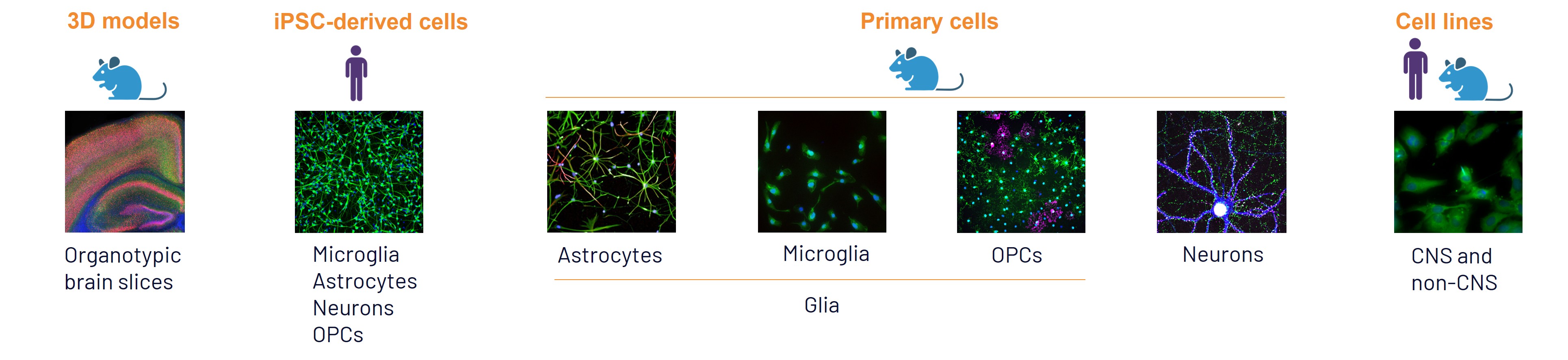

By seamlessly combining rodent and human iPSC-derived models, we deliver robust pre-clinical data for your drug discovery program. Explore our models here.
Discover how human iPSC-derived neural systems, advanced 3D models, and AI-supported workflows are helping improve translational relevance, accelerate neuroscience drug discovery, and enable smarter decision-making for neurological and neurodegenerative disease research in our latest article with Drug Discovery & Development.
Our neuroscience expertise supports drug discovery programs across:
Scientific consultation - We align on target biology, disease mechanism, and program objectives.
Study design and model selection Our scientists recommend the most relevant models, biomarkers, and functional endpoints.
Study execution - Studies are delivered with dedicated scientific oversight and regular communication.
Interpretation and next-step recommendations - Results are translated into actionable conclusions that support progression decisions.
From CNS complexity to confident decisions - Disease-relevant models. Functional biology. Translational biomarkers.
Together, these generate the decision-grade evidence needed to reduce development risk and accelerate neuroscience drug discovery.
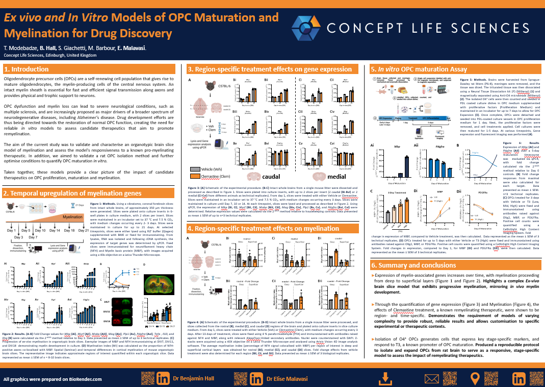
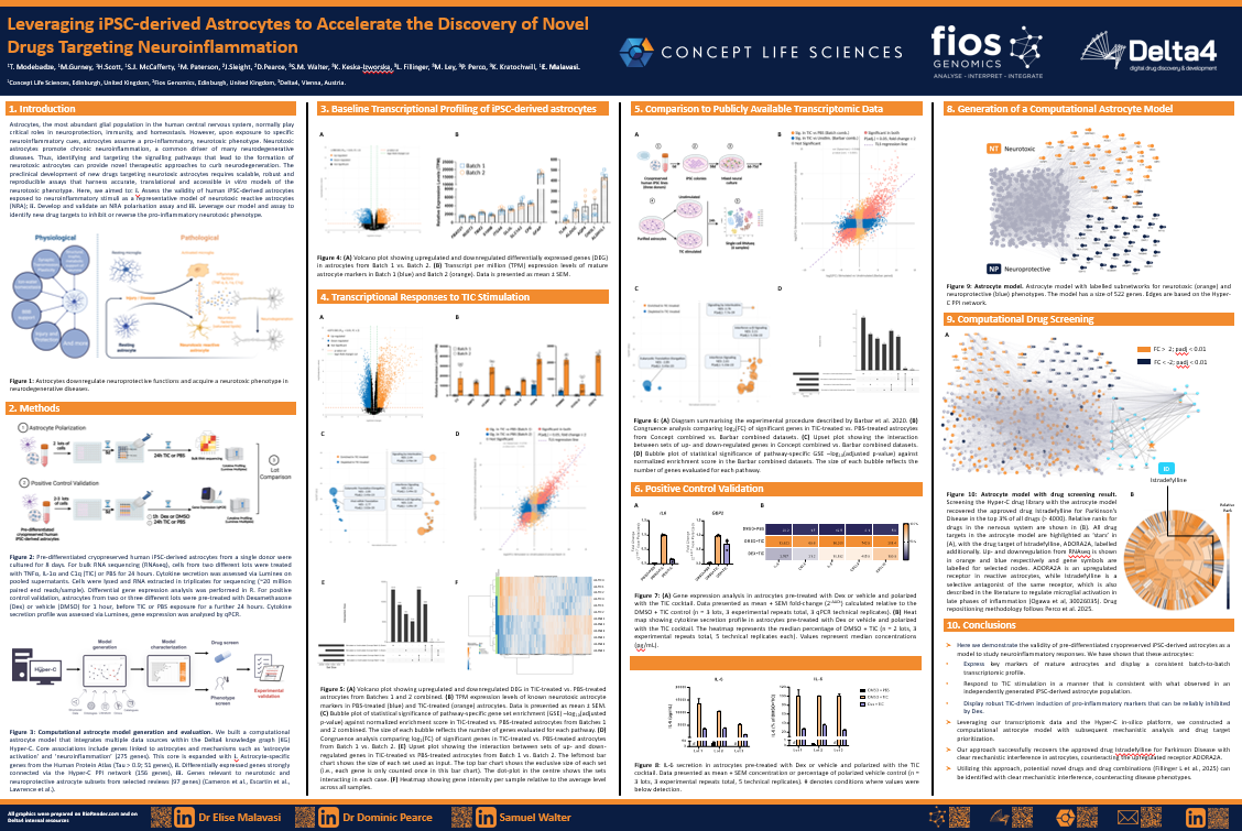
Whether you are validating a novel CNS target, investigating neuroinflammatory mechanisms, evaluating remyelination strategies, or selecting lead candidates, our neuroscience experts can help.
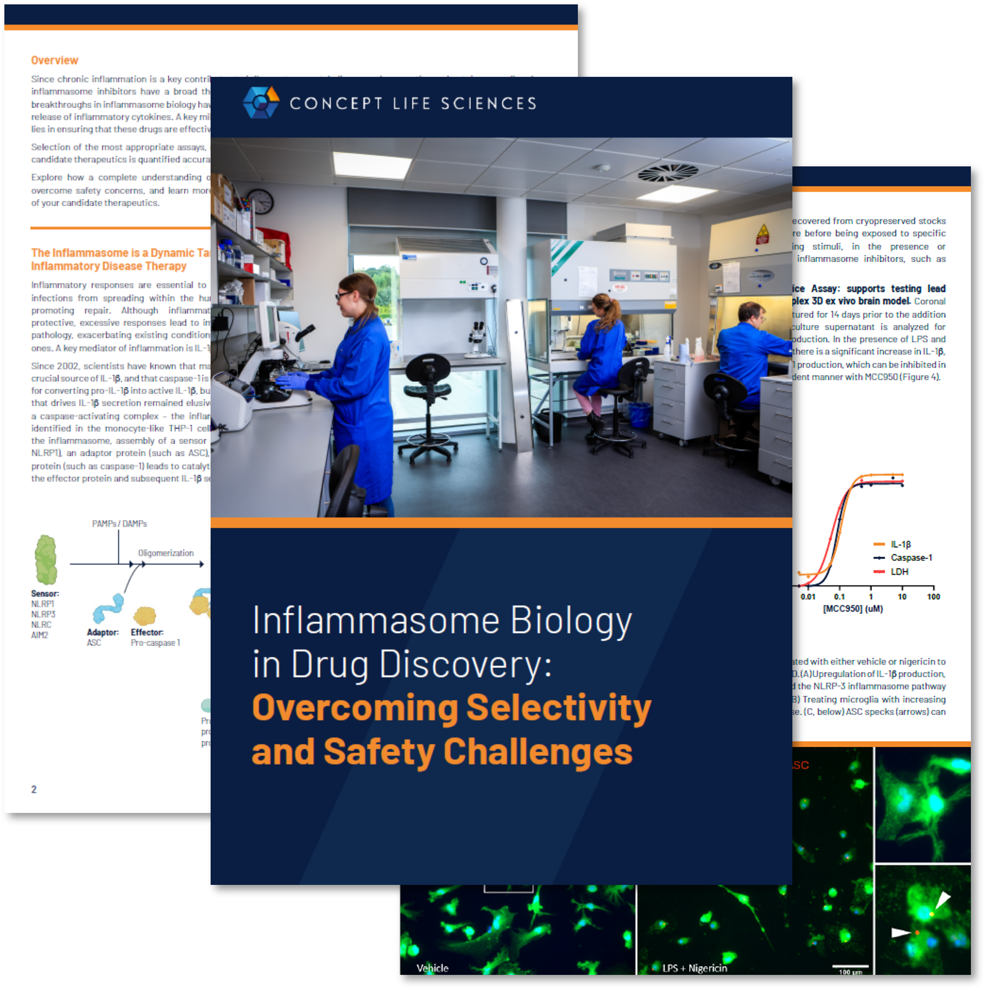
Watch the webinar: Translational Success in Inflammasome Drug Discovery - How advanced models and integrated assays can reduce attrition.
Fast response. Confidential discussions. Direct access to neuroscience experts.
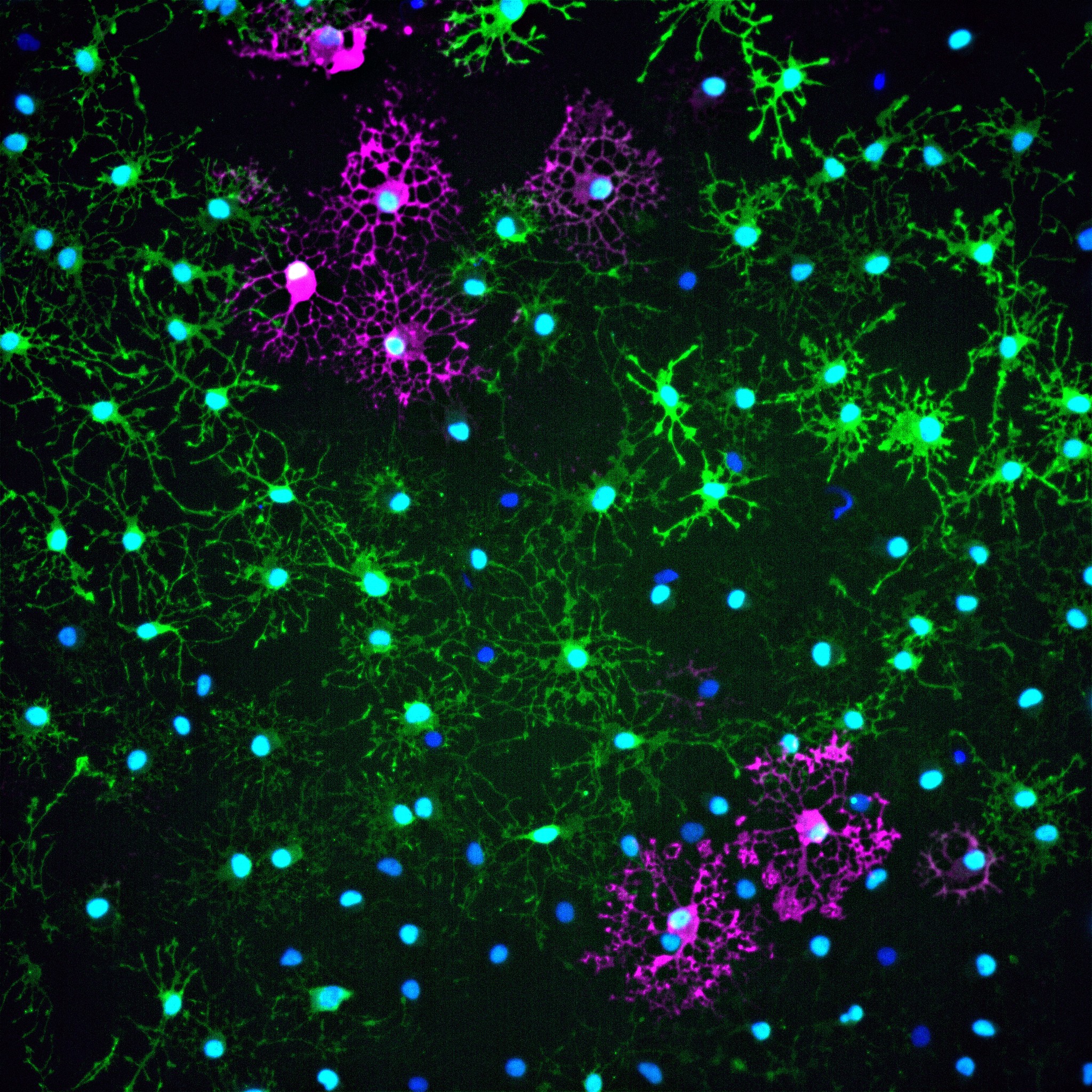
A: We use disease-relevant human and primary CNS systems, including iPSC-derived neurons, astrocytes, microglia, oligodendrocytes, co-culture systems, and organotypic brain slices.
A: Yes. We support drug discovery programs across Alzheimer's disease, Parkinson's disease, ALS, Huntington's disease, multiple sclerosis, and other neurological disorders.
A: We evaluate neuroinflammatory responses using microglial and astrocyte models, cytokine profiling, pathway analysis, and functional cellular endpoints.
A: Yes. We routinely develop and optimize custom assays for novel targets, emerging mechanisms, and complex CNS biology.
A: We combine human-relevant cellular models, functional endpoints, biomarker integration, and mechanism-led study design to improve confidence in clinical translation.