Cell Health and Proliferation Assays
Detect off-target effects and gain data-driven insights

Why is monitoring cell health and proliferation important in drug candidate selection?
Off-target toxicity is one of the leading causes of clinical failure, making cell health and proliferation assays essential for compound screening, toxicology, and therapeutic development. Confidently advance your best candidates with one knowledgeable partner who you can trust to design and run the right assay to support your next decision point.
One partner, end-to-end support
By partnering with Concept Life Sciences, you will benefit from:
- Cost-effective assay design and competitive pricing.
- Confidence at every decision point, from hit-to-lead to candidate selection.
- Access to diverse models, including immortalized, primary, and co-culture systems.
- Tailored readouts for precise interpretation of cellular responses.
Exclude unfit compounds quickly and advance your best candidates with confidence, with our streamlined processes for faster turnaround. All this under one-roof!
Having vast experience in tailored development of in vitro assays, we provide competitive, pre-optimized, ready-to-use solutions, supporting your progression to the clinic. Our integrated approach ensures faster turnaround, reduced risk, and seamless alignment with related in vitro assays such as pathway engagement and ADMET assessments for confident decision making.
Cell painting and advanced live-cell imaging
Cell painting: Morphological fingerprinting for deeper insight
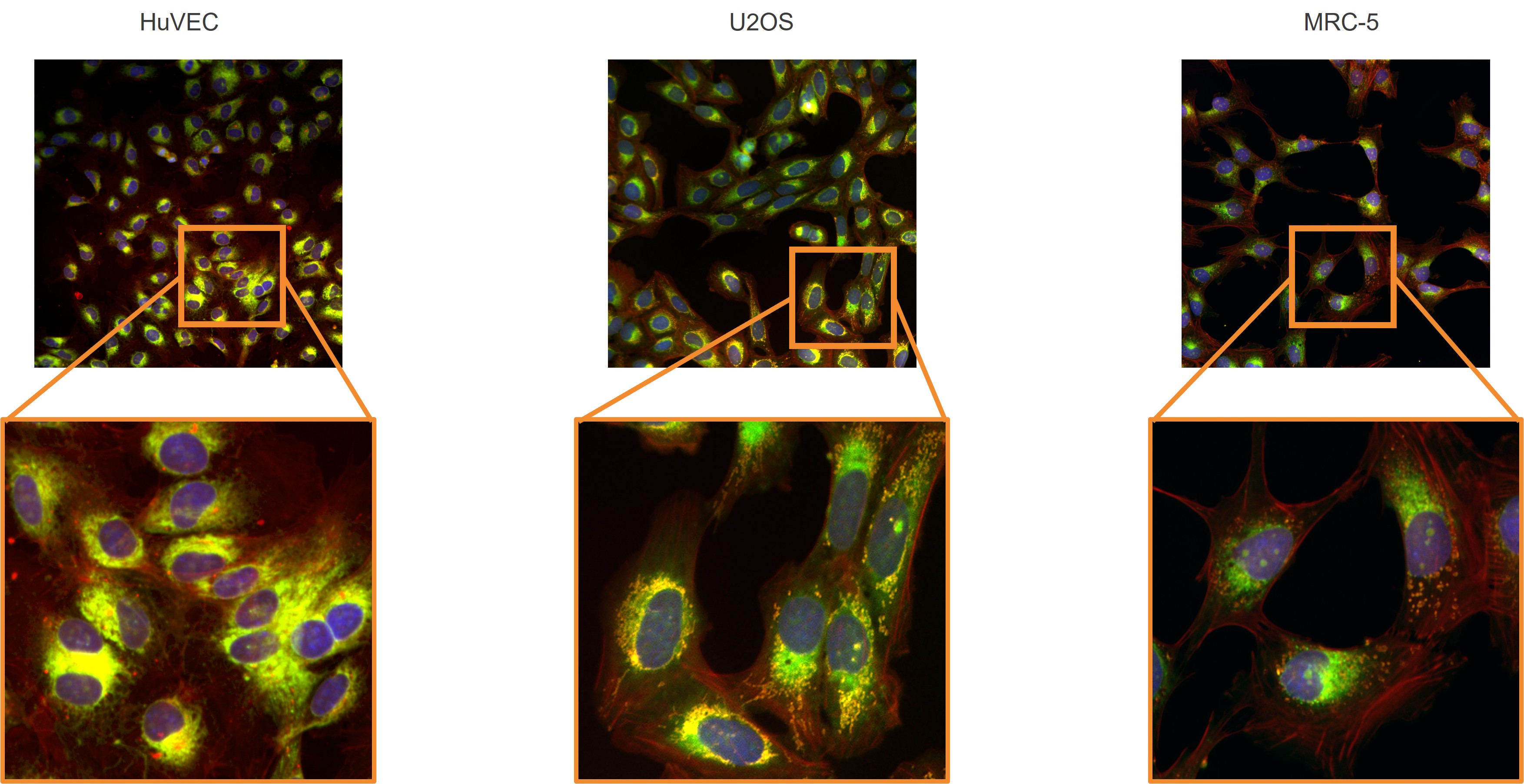
Cell painting is a high-content imaging technique that captures detailed morphological “fingerprints” of cellular health and behavior. This powerful approach helps:
- Predict drug effects and uncover toxicity
- Identify phenotypic changes across multiple cell types
- Differentiate healthy from diseased cells using AI-driven bioinformatics
Unlock the full potential of cell painting across diverse cell types

While U2OS cells are commonly used in cell painting assays, our expertise goes far beyond. We have hands-on experience imaging a wide range of cell types—including immortalized cell lines, iPSC-derived cells, and primary cells. Leveraging our deep knowledge in culturing and imaging diverse cellular models, we offer tailored support to help drive the success of your phenotypic screening projects.
Watch the on-demand webinar - Expanding the cell painting tool kit: Optimizing imaging across diverse and challenging cell lines - where Dr. Andrew Sullivan Principal Scientist, Immunology Concept Life Sciences, explores the challenges and opportunities associated with cell painting, looking at different cell lines and their utility in studying phenotypic changes in response to compounds.
Live-cell imaging with Incucyte®
Leverage the Incucyte® live-cell imaging system for real-time, non-invasive monitoring of cell proliferation, death, and immune response. Designed for high-throughput analysis, this platform provides precise kinetic data to accelerate decision-making and reveal deep biological insights across diverse therapeutic areas.
Comprehensive assays
Our fit-for-purpose, pre-optimized assays are ready to use or fully customizable:
Live cell imaging and morphological monitoring: Visualize and track cell viability and structural changes in real time
- Fluorescent labeling of live cells/nuclei – Non-toxic dyes for real-time imaging.
- High content imaging – Analyze complex morphological changes at single-cell resolution.
- Kinetic monitoring with IncuCyte® – Track cell proliferation and death with a powerful, real-time imaging platform.
Metabolic viability assays: Detect early metabolic changes to determine cellular health
- ATP production – Measure cellular energy status.
- Resazurin reduction – A redox-based indicator of metabolic activity.
- Tetrazolium-based assays (MTT/MTS) – Colorimetric detection of active mitochondrial enzymes.
Plasma membrane integrity: Assess membrane damage and permeability as signs of cell viability or death
- LDH release – Quantify cytoplasmic enzyme leakage into the medium.
- Dye exclusion tests – e.g., Trypan blue or propidium iodide to distinguish live vs. dead cells.
- Membrane permeability assays – e.g., Live/Dead staining kits for fluorescence-based detection
Organelle integrity: Get deeper insights into organelle health, dysfunction, or stress responses
- Mitochondrial membrane potential – Track early apoptotic changes.
- Mitochondrial ROS production – Detect oxidative stress.
- Lysosomal pH – Measure shifts in acidity using pH-sensitive dyes.
- Lysosomal enzyme activity – Assess catabolic function.
- Endoplasmic reticulum (ER) stress – Identify unfolded protein response markers.
Cell death pathway analysis: Characterize specific programmed cell death mechanisms
- Apoptosis – Detect caspase-3 activity and Annexin V binding.
- Necroptosis – Measure RIPK1/RIPK3 phosphorylation.
- Pyroptosis – Quantify caspase-1 activity, IL-1β / IL-18 release, and gasdermin-D cleavage.
Read the case study - Discover how the strategic alliance between Concept Life Sciences and Fios Genomics is advancing complex data analysis in drug discovery — empowering smarter, data-driven decisions.
Cell health and proliferation FAQs
Q: What are cell health and proliferation assays?
A: In vitro assays that measure cell viability, toxicity, and growth to evaluate compound effects during drug discovery.
Q: Why are cell health assays important?
A: They identify off-target toxicity early, preventing costly failures in preclinical and clinical stages.
Q: What technologies are used for live-cell monitoring?
A: Tools such as Incucyte® imaging and cell painting allow real-time, high-content analysis of cellular responses.
Q: Can these assays be customized?
A: Yes. Concept Life Sciences designs tailored assays using the most relevant cell models and detection platforms for your project.
