Peptide profiler
Quick adme quote
Connect with us


Despite promising preclinical findings, many fibrosis drug candidates fail in clinical development due to the limited predictive value of traditional models. A recurring roadblock to successful clinical trials for drugs that target fibrosis is translating positive results in animal models into human disease.
Fibrosis arises from complex interactions between fibroblasts, epithelial cells, immune cells, and the extracellular matrix across multiple organs. Capturing these mechanisms is critical for understanding therapeutic potential.
At Concept Life Sciences, we help bridge this translational gap by crafting innovative customized assays. Using human in vitro cell-based assays, immune-stromal co-cultures, 3D multicellular models, and spatial tissue profiling, we generate mechanistic insights and accurately quantify the effects of candidate drugs that target fibrotic processes, so that you can develop your anti-fibrotic drug programs with confidence.
We support research across multiple fibrosis indications:
Excessive deposition of connective tissue during wound healing can lead to progressive and irreversible damage across multiple organs. As a result, diseases driven by fibrotic pathology—whether in the lung, liver, kidney, skin, or other tissues—are now recognised as major contributors to global morbidity and mortality. Although new drug candidates are emerging, a key challenge remains: how can we adapt cell‑based assays to more accurately model tissue‑specific fibrotic mechanisms and overcome the limitations inherent in animal models?
Concept Life Sciences employs mesenchymal and epithelial cell culture systems derived from a range of relevant tissues to interrogate activation, differentiation and collagen deposition in vitro. This flexibility enables us to mimic fibrosis pathways in organ‑specific contexts. Therapeutics designed to modulate pro‑fibrotic responses can be added at multiple stages of the assays to target distinct phases of fibrosis progression. To further increase physiological relevance, co‑culture systems incorporating accessory cells such as macrophages can be introduced, allowing us to explore immune‑fibroblast crosstalk and better emulate the complexity of tissue microenvironments.
We offer a suite of high-quality cell-based assays to investigate candidate drugs in:
The differentiation of tissue resident mesenchymal cells, such as fibroblasts or pericytes, into myofibroblasts is a key process for effective wound healing. However, it is difficult to assess direct effects of a candidate therapeutic in vivo and animal cell culture does not always translate to clinical success.
To investigate mesenchymal cell differentiation / activation, we culture human fibroblasts and measure changes in viability and gene expression of known activation biomarkers utilizing TGF-β, a recognized pro-fibrotic factor, as a positive control.

High content imaging (HCI) can be used to analyze multiple cellular parameters including collagen production and secretion, cell viability and cell morphology. HCI is suitable for large scale screening studies and provides a robust assessment of phenotypic changes exhibited by mesenchymal cells in response to different treatments.

An additional approach for analyzing collagen type I synthesis is by measuring the secretion of precursor procollagen peptides. Procollagen peptides facilitate the winding of procollagen molecules into a triple-helix and are then cleaved during secretion. The amount of free pro-peptide directly correlates with the synthesis of collagen and can be measured using antibody-based assays. This approach is particularly suited to early screening and efficacy studies
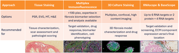
Precise characterization of therapeutic effects within intact tissue architecture is essential for understanding mechanistic activity in fibrotic disease. Our Spatial Biology group employs multiplex immunofluorescence, RNA spatial profiling, and high‑resolution histological imaging to quantify collagen deposition, fibroblast activation states, immune–stromal interactions, and regional heterogeneity within fibrotic lesions. These spatially resolved datasets allow us to track how candidate therapeutics modulate microenvironmental niches, providing a robust framework for mechanistic interpretation and translational prediction beyond conventional in vitro or bulk tissue assays.
Investigating the effects of candidate drugs on human cells is critical to avoid inaccuracies found when translating animal models to human clinical trials. However, selecting disease appropriate cells and assay systems requires expert advice. We offer primary human cell mono-culture assays to confirm potency, efficacy, mode of action and toxicity of accessory cells in vitro. Assay complexity can be increased by co-culturing target cells with mesenchymal cells.
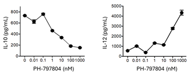
Macrophages are the architects of tissue repair, supporting mesenchymal differentiation, resolving inflammation and promoting tissue repair. However, aberrant macrophage activation leads to fibrosis and immune mediated pathology. Human monocyte derived macrophages can be polarized to disease associated phenotypes and the effect of candidate therapeutics on phenotype and function determined.
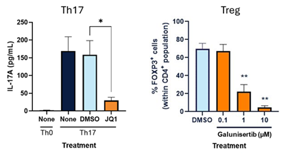
T cells influence fibroblast and macrophage activation through the release of pro- and anti- inflammatory cytokines. Therefore, modifying influential T cell subsets is an important strategy in drug development for fibrotic disease.
We culture primary human T cells with candidate therapeutics to determine whether they alter the inflammatory response. T cells can be polarized to generate disease relevant phenotypes or co-cultured with other cell types of interest.
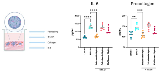
A challenge facing preclinical research is providing assays that imitate the complexity of disease in vivo without relying on animal models. We offer a metabolic associated steatohepatitis (MASH) model that adopts a three-dimensional culture of hepatocytes, stellate cells and macrophages. Cells can be stimulated by lipids in the presence of candidate drugs and changes in cytokine production or pro-collagen determined.
Epithelial to mesenchymal transition (EMT) is a physiological process integral to organogenesis, tissue development, and wound healing. However, it also plays a pathological role in tumor progression, metastasis, and fibrosis. Assays that model epithelial to mesenchymal transition are difficult to find. To investigate EMT during fibrosis, we measure changes in collagen type I and alpha-smooth muscle actin within a scratch wound assay. The assay models the conversion of epithelial cells into fibroblasts and the subsequent wound healing processes, following tissue damage.
Macrophages are pivotal in modulating EMT and a target for novel therapeutics targeting fibrosis. The scratch wound assay can be adapted to include macrophages within the epithelial cell culture supporting analysis of functional changes in co-cultures treated with candidate drugs targeting fibrosis.