Organotypic Brain Slice Inflammasome Assay

Test the efficacy of your novel NLRP3 inhibitors in a physiologically relevant ex vivo system

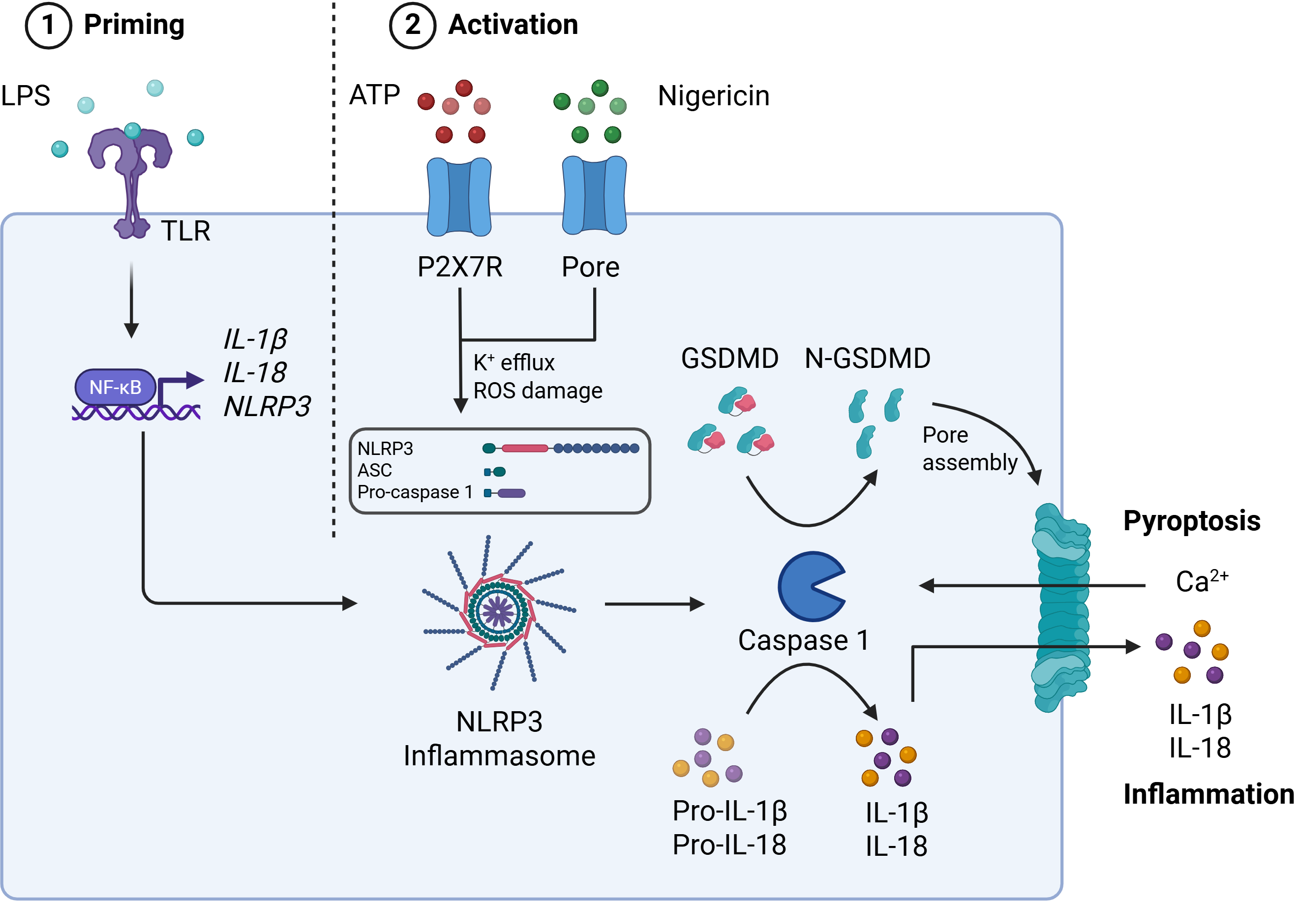
Inflammasomes are large intracellular protein complexes that assemble in response to danger signals and function as key components of the innate immune system. Once activated, they drive Caspase-1–mediated secretion of IL-1β and IL-18 and induce pyroptosis via cleavage of Gasdermin D (Figure 1).

Among inflammasomes, NLRP3 is a well-validated therapeutic target for neurodegenerative disease. It responds to a wide range of damage- and pathogen-associated stimuli, and its chronic activation is implicated in CNS inflammation and pathology.

Figure 1. NLRP3 activation requires a priming stimulus and an activation stimulus. The priming step (e.g. via LPS-induced NF-κB signaling) induces NLRP3, and pro-IL-1β expression and pro-IL-18 expression. The second step involves endogenous (e.g. ATP) or exogenous (e.g. Nigericin) signals that drive cytosolic assembly of NLRP3, ASC, and Caspase-1. Activated Caspase-1 cleaves pro-IL-1β, pro-IL-18, and Gasdermin D—the latter forming membrane pores, releasing cytokines and triggering pyroptosis.

Key benefits

Our brain slice inflammasome assay provides a powerful 3D ex vivo platform to assess efficacy of NLRP3 inhibitors in a native CNS environment.

  • Preserves key CNS cell populations and cytoarchitecture for physiologically relevant insights
  • Enables robust, reproducible readouts of inflammasome activation and inhibition
  • Supports a range of end-point analyses including ELISA, western blot, immunohistochemistry, qPCR, and RNAseq
  • Cost-effective and faster than in vivo models
  • Adaptable to other inflammasome pathways and disease mechanisms

Protocol

Figure 2.Brain slice preparation and stimulation protocol. Coronal forebrain slices are prepared from neonatal C57BL/6 mouse brains using a vibratome and cultured on inserts. After a 14-day maturation period, slices are primed with LPS and treated with vehicle or the NLRP3 inhibitor MCC950. Activation is induced using ATP or Nigericin. Supernatants are then collected for cytokine analysis.
Compound requirements
Test systems
Assay format
Controls
Quantitation
Deliverables

Data analysis & results

Selective activation of the NLRP3 inflammasome with ATP or Nigericin induces robust IL-1β secretion

Figure 3. Secreted levels of IL-1β in tissue culture supernatants from slices treated with either LPS alone, LPS + ATP (A) or LPS + Nigericin (B), either in the absence or presence of NLRP3 inhibitor MCC950.

The secretion of multiple NLRP3 activation markers is concentration-dependently inhibited by MCC950

Figure 4. Secreted levels of IL-1β (A), Gasdermin D (B), HMGB1 (C) in tissue culture supernatants from slices treated with LPS + ATP, either in the absence or presence of increasing concentrations of NLRP3 inhibitor MCC950.

After supernatant collection, brain slices can be utilized for various end-point readouts, including western blotting, immunohistochemistry, RTqPCR, and RNAseq

Figure 5. Wide field fluorescence image of a coronal organotypic brain slice stained with antibodies to Neurofilament heavy chain (green), NeuN (red), and counterstained with DAPI (blue). Image acquired with Vectra PolarisTM Imaging System.

Conclusions

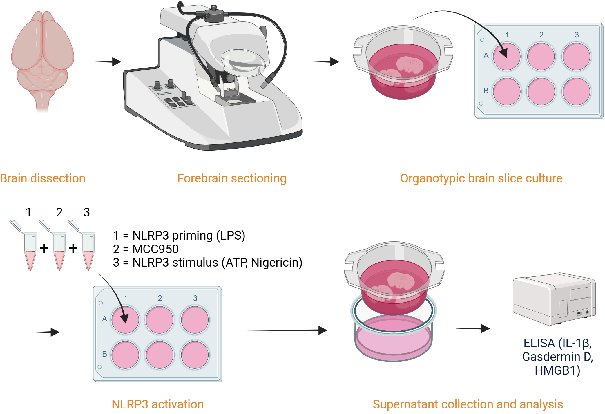
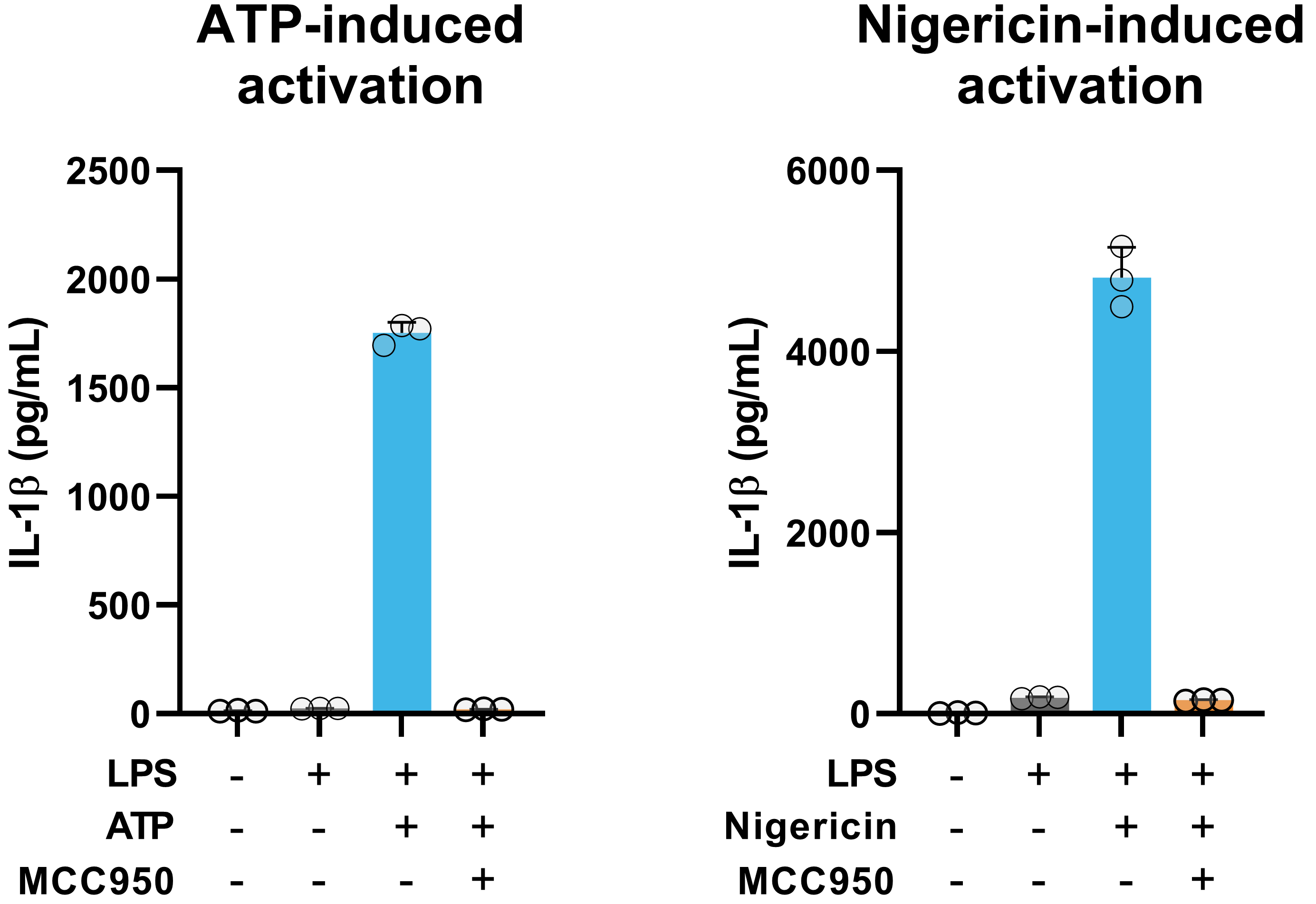
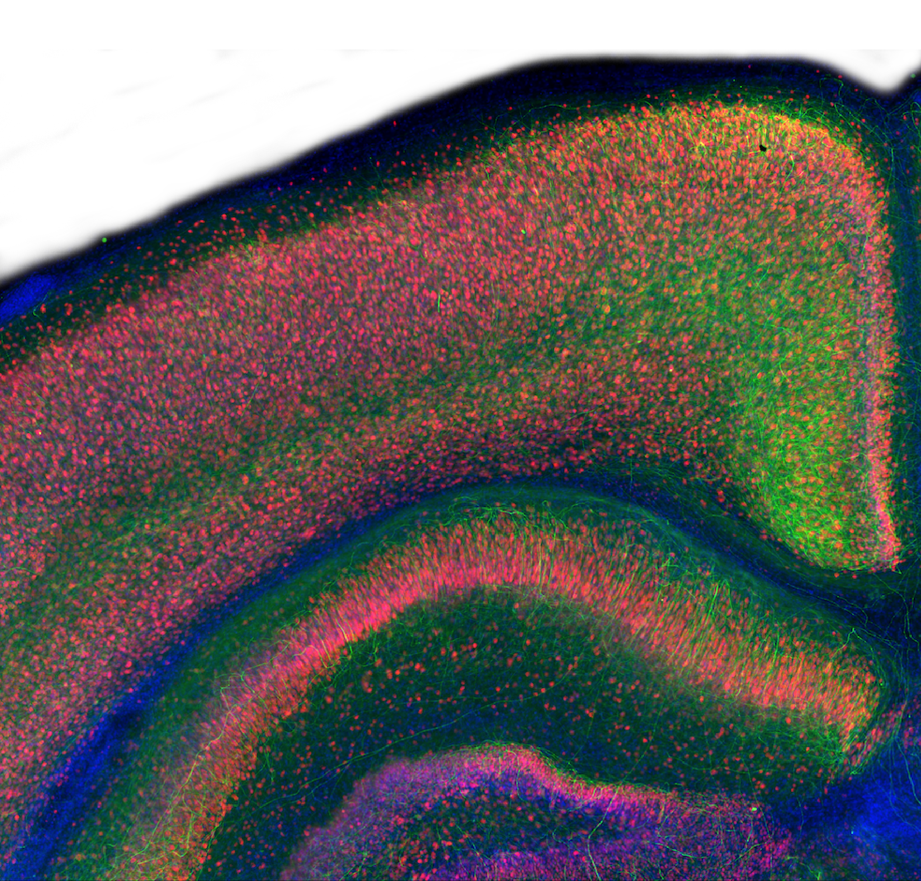
NLRP3 activation contributes to the neuroinflammation that drives neurodegenerative diseases such as Alzheimer’s and Parkinson’s. Our organotypic brain slice assay captures key aspects of CNS inflammasome biology, enabling accurate assessment of NLRP3 inhibitors in a complex and native tissue environment.

By offering this physiologically relevant platform, we support in-depth compound profiling and mechanism-of-action studies; de-risking your in vivo studies and helping you progress promising candidates with confidence.