We connect you with specific subject matter experts to ensure support and recommendations tailored to your project.

Thank you, your request has been received.

We're busy connecting you with an expert, who will be in touch as soon as possible.

Oops! Something went wrong while submitting the form. Please try again or email us at webenquiries@conceptlifesciences.com
We will respond to you within 24 hours of your enquiry being received.

You can also find Concept Life Sciences on the following platforms:

Human iPSC-derived Microglia Inflammasome Assay

Confirm the efficacy of brain-penetrant candidate NLRP3 inhibitors in human iPSC-derived microglia

Why target the NLRP3 inflammasome in the brain?

Inflammasomes are large intracellular protein complexes that assemble in response to danger signals and function as key components of the innate immune system. Once activated, they drive Caspase-1–mediated secretion of IL-1β and IL-18 and induce pyroptosis via cleavage of Gasdermin D (Figure 1).

Among the inflammasomes, NLRP3 is a well-validated therapeutic target for neurodegenerative disease because its chronic activation is implicated in CNS inflammation and pathology.
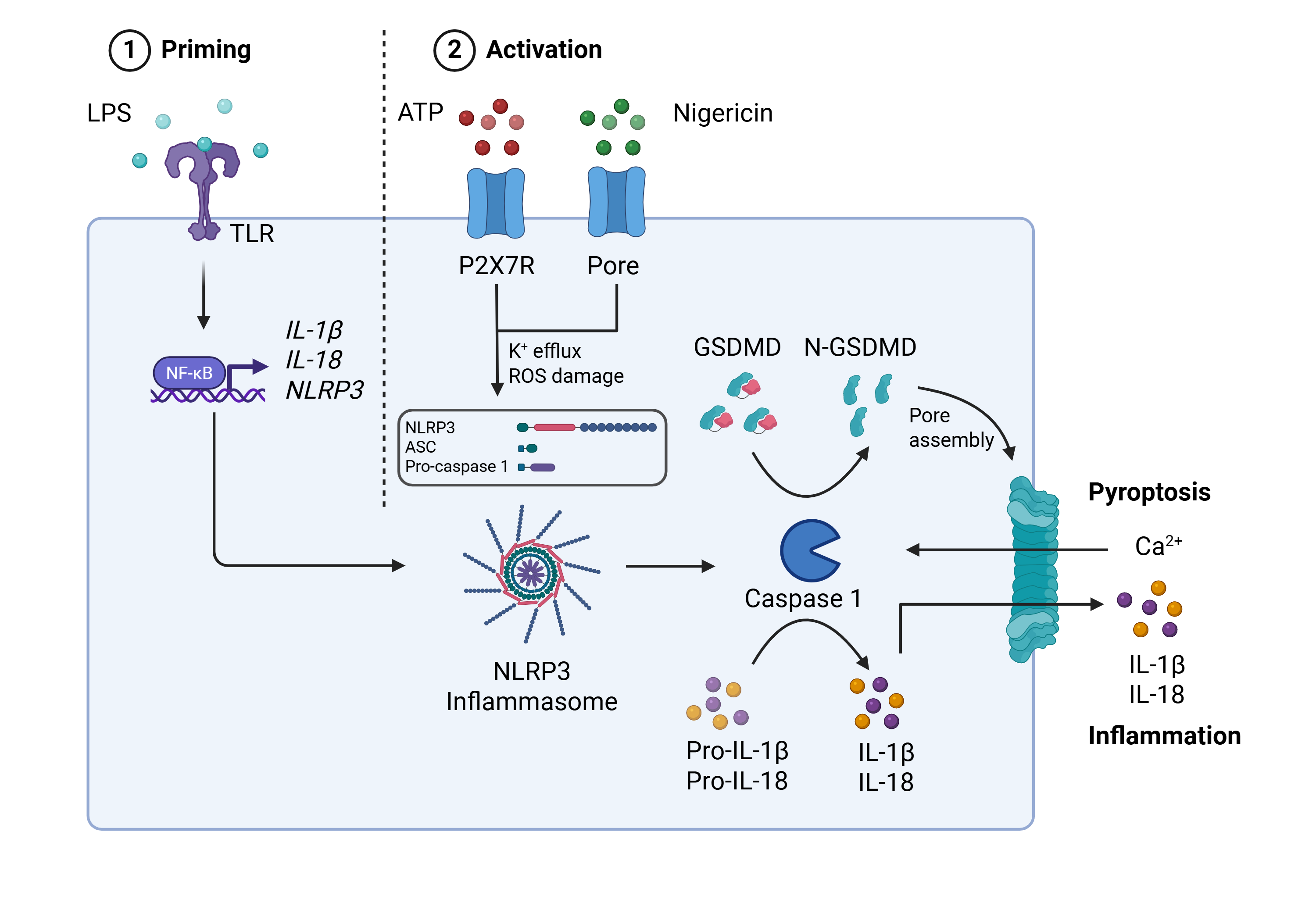
Figure 1. NLRP3 activation requires a priming stimulus and an activation stimulus. The priming step (e.g. via LPS-induced NF-κB signalling) induces NLRP3, and pro-IL-1β expression and pro-IL-18 expression. The second step involves endogenous (e.g. ATP) or exogenous (e.g. Nigericin) signals that drive cytosolic assembly of NLRP3, ASC, and Caspase-1. Activated Caspase-1cleaves pro-IL-1β, pro-IL-18, and Gasdermin D—the latter forming membrane pores, releasing cytokines and triggering pyroptosis.

Challenges in NLRP3 drug discovery

Drug developers must overcome several hurdles to advance brain-targeting NLRP3 inhibitors to the clinic, including:

Uncertain CNS target engagement: CNS-permeant NLRP3 inhibitors may look promising in biochemical or cell-based high throughput screening assays, but fail to show meaningful activity in human microglia, where the therapeutic mechanism matters.

Poor translation: Rodent or surrogate myeloid systems may not predict human microglial biology well enough for decision-making.

Selectivity and off-target risk: NLRP3 programs can be confounded by nonspecific anti-inflammatory effects or toxicity.

Lack of robust biomarkers: Robust human pharmacodynamic evidence is needed before advancing compounds into more expensive in vivo and clinical studies.

Key benefits

The assay addresses the key risks in NLRP3 drug discovery by confirming that brain-penetrant candidates are not only reaching the CNS, but also functionally inhibiting inflammasome activation in human microglia, the cell type most directly linked to neuroinflammatory pathology.

  • Physiological relevance for CNS indications: Test compound efficacy in human microglia, the key drivers of neuroinflammation in the CNS.
  • Disease relevance: Assess compound activity in microglia carrying relevant disease-associated mutations.
  • Target engagement and potency: Use the industry-standard Caspase-1 activation readout to confirm inflammasome inhibition and measure drug potency.
  • Built-in orthogonality: Increase confidence by measuring IL-1β release, LDH release, and ASC speck formation alongside Caspase-1 activity.

Protocol

The key differentiators:

Orthogonal confirmation: Multiplex up to 4 readouts with different modalities, maximizing efficiency and insight, while minimizing the risk of assay artifacts.

Physiological relevance: Assess candidates against the endogenously expressed target in the relevant cell type, eliminating the risk of overexpression-induced artifacts and minimizing the risk of poor translation.

Platform integration: Combine our iPSC microglia NLRP3 assay with our THP-1 NLRP3 screening assay and our Organotypic Brain slice NLRP3 assay to build a screening cascade.
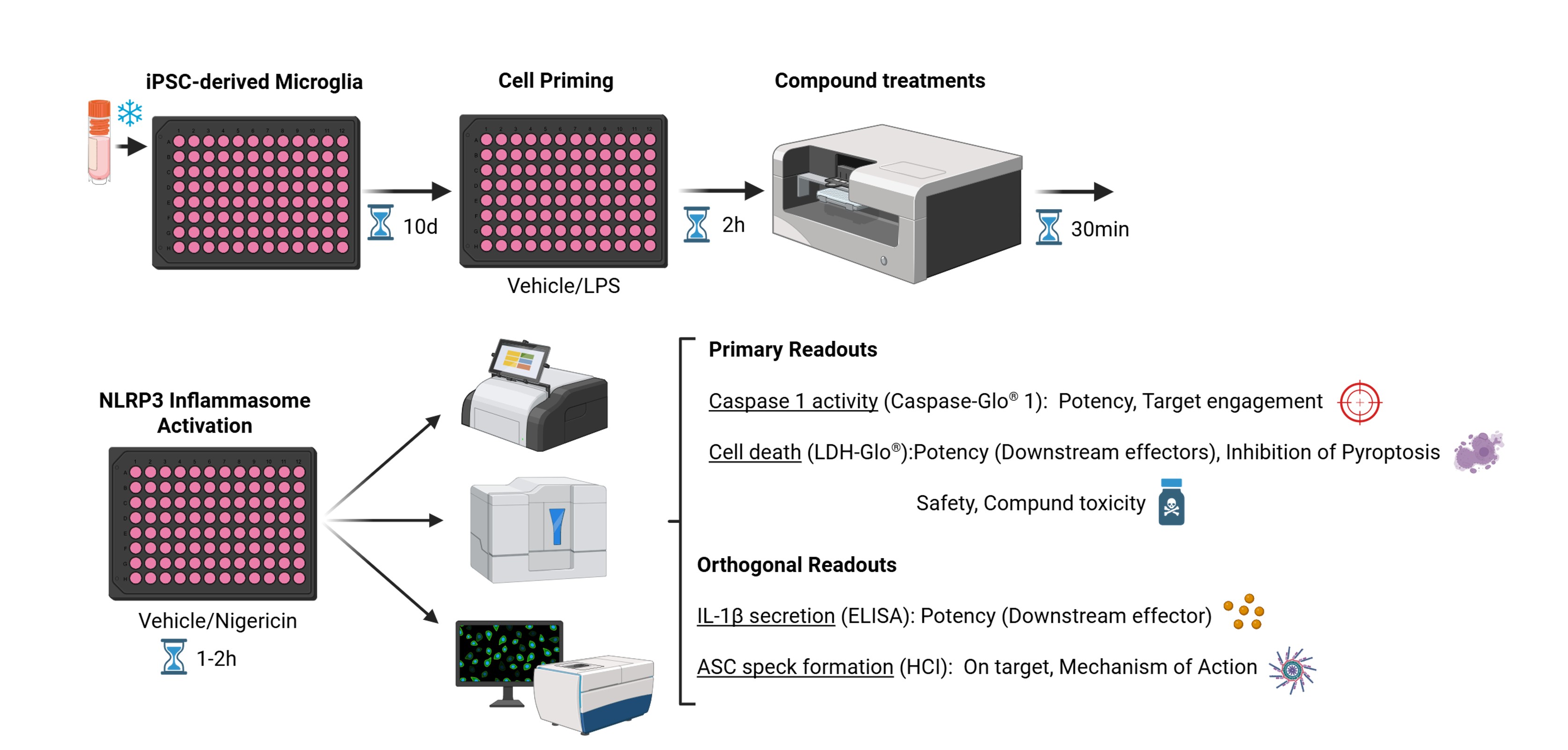
Figure 2. NLRP3 stimulation protocol. Human iPSC-derived microglia (microglia) are recovered from cryopreserved stocks and stabilized in culture for 10 days before being primed with either vehicle or LPS for 2 hours. Next, the cells are pre-treated with test compounds for 30 minutes, followed by NLRP3 activation with either vehicle or Nigericin for up to 2 hours. Cell culture supernatants are collected and used to quantify Caspase-1 activity and LDH release, with optional confirmation of IL-1β concentration. The cells are fixed and stained with a fluorescently labelled anti-ASC antibody and CellMaskTM, with nuclei counterstained with DAPI. Stained cells are analyzed by High Content Imaging.
Compound requirements
Test systems
Assay format
Controls
Quantitation
Deliverables

Data analysis and results

LPS priming followed by Nigericin induces strong NLRP3 inflammasome activation in microglia.
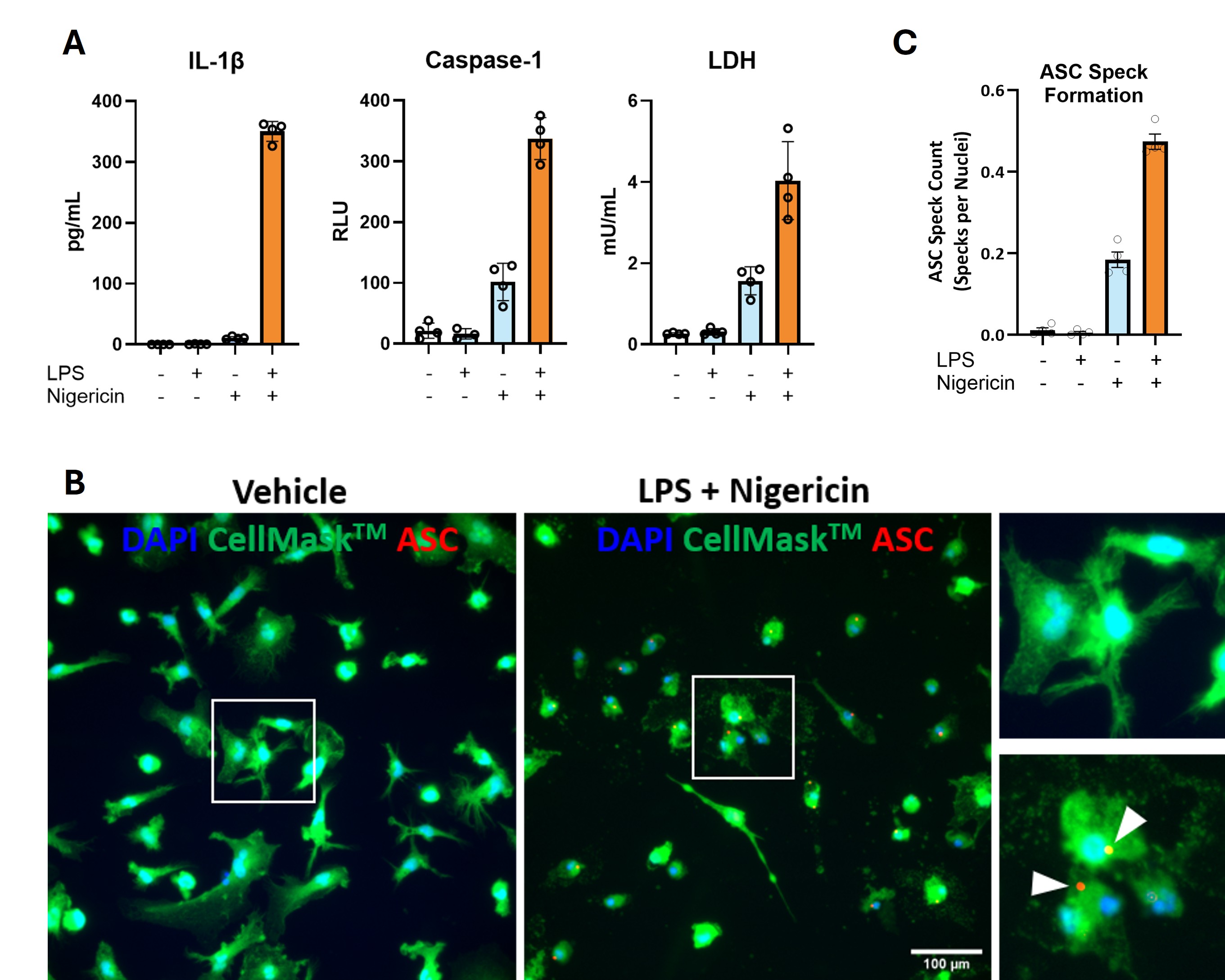
Figure 3. (A) IL-1ꞵ, Caspase-1 activity and LDH (an indicator of pyroptosis) were quantified in cell culture supernatants from microglia exposed to the indicated stimuli. (B) Microglia were immunostained with a fluorescently labelled anti-ASC antibody (red), CellMaskTM (green), and DAPI (blue) after treatment with vehicle or LPS + Nigericin. Arrowheads point to ASC specks. (C) ASC specks were quantified by High Content Imaging in microglia exposed to the indicated stimuli. All data shown represents the mean (± SEM) of four technical replicates.

The selective NLRP3 inhibitor MCC950 induces concentration-dependent inhibition of microglial inflammasome activation.
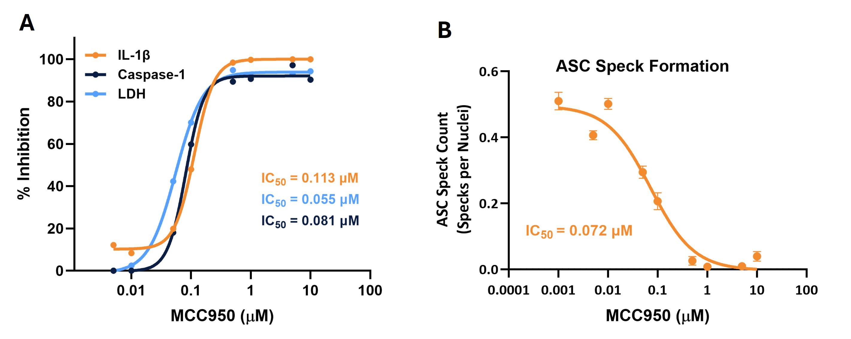
Figure 4. (A) Positive control validation. Concentration-dependent inhibition of IL-1ꞵ release, Capase-1 activity and LDH by reference NLRP3 inhibitor MCC950. The calculated IC50 is indicated for each readout. (B) ASC specks were immunolabelled and quantified by High Content Imaging in microglia stimulated with LPS and Nigericin in the presence or absence of increasing concentrations of NLRP3 inhibitor MCC950. The calculated IC50 is shown.

Conclusions

NLRP3 inflammasome activation is not only a consequence of CNS tissue injury in neurodegenerative diseases but also a driver of the neuroinflammatory cascades that fuel their pathogenesis. Therefore, CNS-targeted NLRP3 inhibitors may offer therapeutic benefit across multiple neurodegenerative conditions, such as Alzheimer’s and Parkinson’s disease.

Our human iPSC-derived microglia inflammasome assay delivers robust functional data on candidate CNS-targeting NLRP3 inhibitors, giving you decision-ready human evidence to confidently de-risk your progression into the clinic.