Peptide profiler
Quick adme quote
Connect with us


Our experienced immuno-oncology team works with advanced human cell models to provide physiologically relevant insights that accelerate your drug discovery. As the first CRO to develop and offer a comprehensive suite of tumor-associated macrophage (TAM) and exhausted T cell assays a decade ago, we’ve supported over 100 immuno-oncology drug discovery programs to date.
Immuno-oncology has revolutionized cancer treatment, with first-generation checkpoint inhibitors showing strong results in advanced cancers. However, resistance mechanisms can limit their effectiveness. As the field advances, focus is shifting towards broader immune-modulating strategies, highlighting the need for sophisticated tools to decode complex tumor-immune interactions.
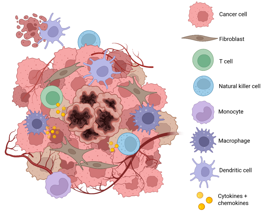
At Concept Life Sciences, we offer cutting-edge in vitro assay platforms that faithfully model the tumor microenvironment. These systems capture the intricate interplay between cancer cells, stromal components, and immune modulators, providing a powerful foundation for identifying and validating novel therapeutic targets.
Whether you're exploring new immunotherapy candidates or refining existing strategies, our assays deliver biologically relevant insights that accelerate discovery and de-risk development.
With deep expertise rooted in our legacy as Aquila Biomedical, we’ve been at the forefront of translational immuno-oncology for nearly a decade. As the first CRO to develop and offer a comprehensive suite of tumor-associated macrophage (TAM) and exhausted T cell assays, we’ve supported over 100 immuno-oncology drug discovery programs to date.
Our assays deliver critical, decision-enabling data that expands understanding of target and compound behaviour, provides robust experimental validation of client hypotheses, and helps unlock the next stages of funding and development.
Whether you're advancing early-stage discovery or preparing for clinical translation, our platforms are designed to accelerate progress and empower confident decision-making.
Accelerate your immuno-oncology program with confidence using advanced human-relevant in vitro models designed to better predict clinical outcomes.
Human biology at the core - Our assays are built on primary human immune cells—PBMCs, isolated T cells, and other immune subsets—offering a more accurate representation of patient responses than traditional models.
Expertly engineered by immunologists and cell biologists - We’ve developed a suite of robust, reproducible assays that capture key pathological processes in cancer, including immune suppression, T cell exhaustion, and stromal cell interactions.
Versatile platforms for diverse needs - From monoculture to co-culture, 2D to 3D spheroid models, we support a wide range of therapeutic modalities. Our assays are available off-the-shelf or can be tailored to your specific program.
CRO-leading expertise in complex phenotypes - We specialize in modelling tumor-associated macrophages, cancer-associated fibroblasts, and exhausted T cells—delivering more faithful representations of the tumor microenvironment without the variability of patient-derived cells.
At the forefront of immuno-oncology innovation, we offer a comprehensive suite of cutting-edge assays designed to support the discovery and development of next-generation cancer immunotherapies. Our platforms are tailored to address the most promising therapeutic strategies, including:
Explore our featured assays below:
We develop advanced T cell assays that replicate functionally distinct, exhausted T cell states commonly observed in cancer patients. These models are ideal for studying the mechanisms of T cell exhaustion, identifying novel immunotherapy targets, and evaluating compound efficacy in reversing dysfunctional T cell responses.
Our co-culture assays are designed to assess how compounds influence the reprogramming of TAMs and their downstream effects on T cell suppression. These models help elucidate the immunosuppressive tumor microenvironment and support the development of strategies to restore effective anti-tumor immunity.
We build complex 3D assay systems incorporating cancer cells, cancer-associated fibroblasts, and immune cells to better mimic the tumor microenvironment. These models provide a more physiologically relevant platform for testing compound activity, enabling deeper insights into therapeutic potential and translational relevance.
Whether you're in early discovery or late-stage validation, our robust, customizable assay platforms provide the insights you need to accelerate your immuno-oncology pipeline.
Our specialist immuno-oncology translational biology expertise is complemented by a wider offering across chemistry, ADMET, and GMP manufacturing services. Speak to our team to explore how we can help you bring your immunology therapeutic to the clinic.


