Peptide profiler
Quick adme quote
Connect with us


Our specialist Immunology team combines decades of experience in immunology with advanced human cell models to provide physiologically relevant insights that accelerate your drug discovery. With additional services across chemistry, ADMET and GMP manufacturing, we are able to provide seamless support across the drug discovery and development pipeline.
Watch our new webinar: Translational Success in Inflammasome Drug Discovery - How advanced models and integrated assays can reduce attrition.
Immunology is inherently complex, demanding deep expertise and specialized solutions to translate research into real-world impact. Our expert team delivers high-quality data and insight to empower decision-making throughout drug discovery and development. By using advanced human immune cell models, we generate physiologically relevant data that enhances the translational success of your program.
With a legacy spanning over 15 years, we pioneered translational immunology assays as Aquila Biomedical, and since then have continued to support clients as they bring cutting-edge immunology therapies to the clinic.
With our flexible and consultative approach, we work closely with you to provide custom-built solutions that are led by your goals, timelines and internal processes to deliver the insight needed to drive your project to the next stage.
Our suite of assays include:
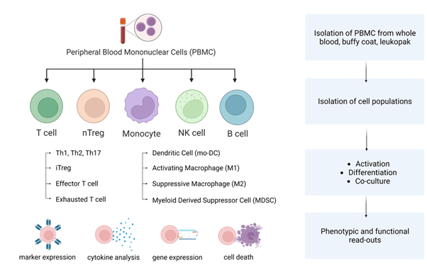
Human models are essential to more accurately predict human immune responses and clinical efficacy. Our team of immunologists has developed robust in vitro assays to recapitulate many key pathological processes relevant to the immunotherapy field. Our assays use human primary immune cells for the closest representation of clinical settings, delivering the most meaningful pre-clinical data for your program of work.
We have significant experience working with human primary immune cells, either whole PBMC, isolated T cells or other immune cells to assess compound effects in monoculture or co-culture assay systems, 2D and 3D spheroid models, and across many different modalities.
In addition, we offer readout to support your objectives, including comprehensive high-dimensional readout such as transcriptomics, high-content imaging and multiplex assays.
Discover how human iPSC-derived neural systems, advanced 3D models, and AI-supported workflows are helping improve translational relevance, accelerate neuroscience drug discovery, and enable smarter decision-making for neurological and neurodegenerative disease research in our latest article with Drug Discovery & Development.
Our team brings over 10 years of experience in developing assays that model key aspects of immune cell interactions with cancer, supporting both mechanistic studies and therapeutic development.
We develop advanced T cell assays that replicate functionally distinct, exhausted T cell states commonly observed in cancer patients. These models are ideal for studying the mechanisms of T cell exhaustion, identifying novel immunotherapy targets, and evaluating compound efficacy in reversing dysfunctional T cell responses.
Our co-culture assays are designed to assess how compounds influence the reprogramming of TAMs and their downstream effects on T cell suppression. These models help elucidate the immunosuppressive tumor microenvironment and support the development of strategies to restore effective anti-tumor immunity.
We build complex 3D assay systems incorporating cancer cells, cancer-associated fibroblasts, and immune cells to better mimic the tumor microenvironment. These models provide a more physiologically relevant platform for testing compound activity, enabling deeper insights into therapeutic potential and translational relevance.
Our team develops specialized in vitro models to investigate immune mechanisms underlying autoimmune diseases and evaluate the therapeutic potential of novel compounds.
We use isolated B cells from PBMCs and stimulate them with disease-relevant agents to model B cell hyperactivity seen in autoimmune conditions. These assays allow assessment of a compound’s ability to suppress B cell activation, with readouts including proliferation, activation marker expression, differentiation, and antibody production.
These assays evaluate how compounds influence dendritic cell maturation and their capacity to activate antigen-specific T cells. Readouts include maturation marker expression and cytokine release, providing insight into a compound’s potential to dampen immune activation at the antigen presentation stage.
These models support mechanistic studies and compound screening in the context of immune tolerance, inflammation, and autoimmune pathogenesis, helping researchers better understand therapeutic mechanisms and refine development strategies.
We offer a range of well-characterized assays to model key inflammatory processes, enabling detailed evaluation of compound effects on immune cell function and inflammatory signalling.
We model T cell polarization under defined culture conditions to generate TH1, TH2, and TH17 phenotypes, which are commonly associated with various inflammatory disorders. These assays are used to assess a compound’s ability to inhibit or modulate T cell differentiation, providing insight into potential anti-inflammatory mechanisms.
Using a panel of functionally distinct macrophage subtypes, we evaluate how compounds influence macrophage polarization, particularly their shift away from pro-inflammatory or disease-associated phenotypes. This supports studies focused on immune resolution and tissue repair.
We offer a suite of assays using THP-1 cells and primary macrophages to assess compound activity on the inflammasome pathway. Readouts include ASC speck formation, IL-1β release, and other markers of inflammasome activation, enabling detailed profiling of anti-inflammatory potential.
Our specialist immunology translational biology expertise is complemented by a wider offering across chemistry, ADMET, and GMP manufacturing services. Speak to our team to explore how we can help you bring your immunology therapeutic to the clinic.


