Peptide profiler
Quick adme quote
Connect with us


We believe great science starts with effective scientific partnership. That’s why our biology services are built on collaboration, combining deep scientific expertise, flexibility and seamless integration across disciplines to address your unique challenges and move your discovery forward faster, with confidence.
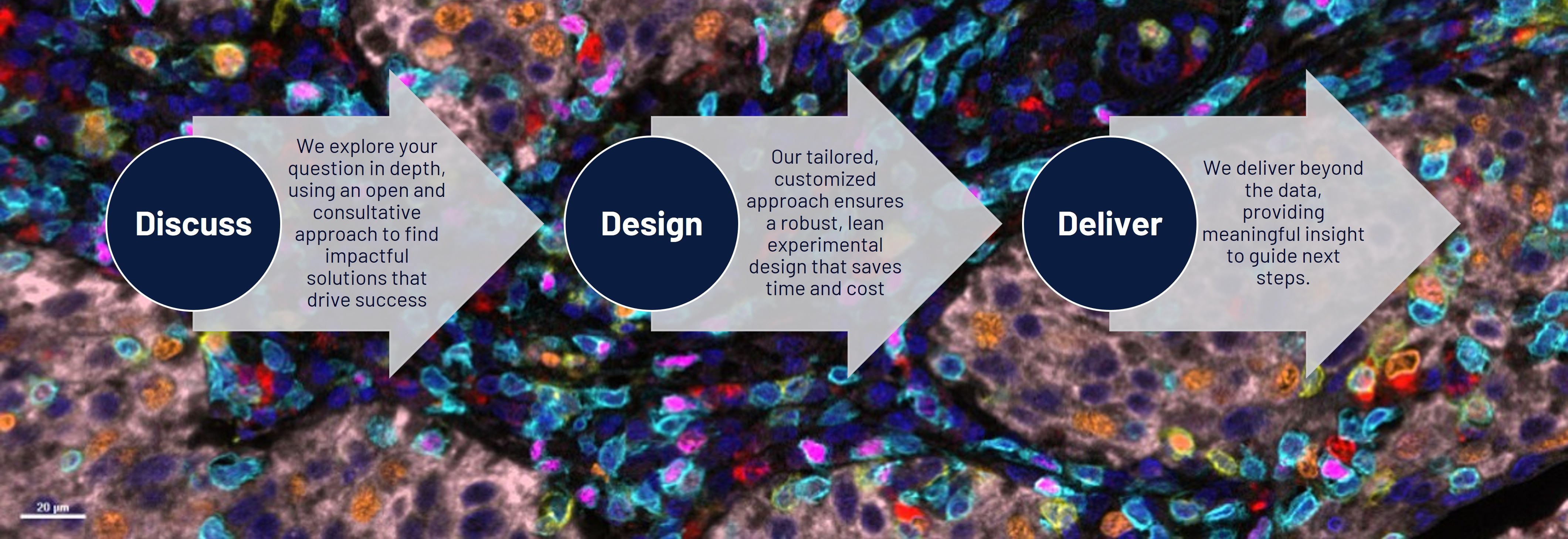
The biology behind drug discovery is more complex than ever: novel modalities, limited budgets, tight timelines, and scattered outsourcing can slow progress. Our Discuss-Design-Deliver framework is powered by deep translational biology expertise to find the best solution first time, getting you from target to optimized lead faster. By implementing the fail-fast approach the focus is only on those assets with demonstrable efficacy and /or the right efficacy profile, resulting in fewer detours and greater confidence in decision-making.
We understand:
We understand these pressures because we’ve lived them through our decades of experience using advanced human-relevant systems. Our mission is to simplify your journey from idea to insight by building a biology partnership that feels like an extension of your team.
Integration across discovery: Our biosciences, chemistry, and translational experts collaborate in real-time, for an efficient and iterative drug discovery process.
Assay development and screening: Robust, predictive assays and optimized screening strategies accelerate hit-to-lead progression and reduce risk. From tailored assay design to complex profiling, delivering actionable insights.
Disease modeling: Leveraging physiologically relevant models, from primary cells and iPSCs to complex co-culture systems, that capture disease complexity and enable more accurate predictions for better translational outcomes.
Advanced models and spatial biology: From 3D in vitro and ex vivo models to spatial biology, histology and high-content imaging, our platforms deliver data that capture biological complexity.
Customisable assay design: Built around your science because every project is unique. We design assays tailored to your target, readout, and mechanism, from small molecules to gene and cell therapies.
Breadth of expertise across modalities and therapy areas: from small molecules to cell and gene therapies for confidence in predictive insights across oncology, immunology, neuroscience, fibrosis, and more.
Insightful data that drives action: Our expert scientists don’t stop at data generation, they interpret your results, highlight key insights and recommend next steps.
Transparent communication: Responsive, flexible and invested in your success, our teams are an extension of your team. As one of our biotech clients says: “Concept Life Sciences feels like an extension of our own team, scientifically sharp, fast, and collaborative.”
Integration. Expertise. Insight.
We go beyond generating data, we interpret it, connect it and help you act on it.
When you work with us, you’re not choosing another CRO, you’re choosing a team that’s invested in your success and aligned with your goals.
Scientists first and collaborators always.
Start your next biology project with confidence
Work with scientists who care as much about your program as you do. Whether you’re exploring a new target or refining translational models, we’ll help you design, test and interpret biology that matters.


